Purpose
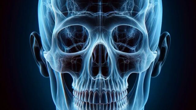
Observation of trauma and lesions in the vicinity of the skull base, conical bone, oval foramen, spinous foramen, sphenoidal sinus, ethmoidal sinuses, etc.
Useful in facilities without CT equipment.
Prior confirmation
In the case of trauma patients, confirm the absence of cervical dislocation, fractures, or suspected spinal cord injuries in the cervical spine.
Verify the purpose of the examination.
Remove any obstructing objects (hairpins, glasses, earrings, necklaces, dentures, etc.)
Positioning
Position the patient in a seated position with their back against the imaging table (supine position is also acceptable).
Note: The probability of experiencing dizziness is higher in the supine position.
Align the mid-sagittal plane and the cassette perpendicularly.
Place markers (R or L) accordingly.
Elevate the chin and align the Frankfurt Horizontal plane parallel to the cassette.
CR, distance, field size
CR : Align the crosshairs (vertical) of the radiation field with the mid-sagittal plane and position the point where the crosshairs (horizontal) of the radiation field pass through the external auditory meatus at a caudal direction with a 10° oblique angle.
Distance : 100cm.
Field size : Include the entire head, including soft tissues.
Exposure condition
75kV / 32mAs
Grid ( + )
Suspend respiration.
Image, check-point
Normal (Radiopaedia)
Ensure that the chin is positioned anterior to the sphenoidal sinuses.
Confirm that the zygomatic arches are projected symmetrically on both sides.
Verify that the mandible is projected symmetrically on both sides.
Ensure that the entire skull is included in the image.
Ensure there is no motion blur.
Confirm the visibility of the sphenoidal sinuses and ethmoidal sinuses.
Videos
Related materials


