Purpose
Observation of fractures, dislocations, lesions, and trauma to the humerus.
Presence or absence of osteoporosis.
Prior confirmation
Confirm whether the patient is to be radiographed in the standing (seated) or supine position.
If a fracture is suspected, do not move the arm.
Remove any obstacles.
Positioning
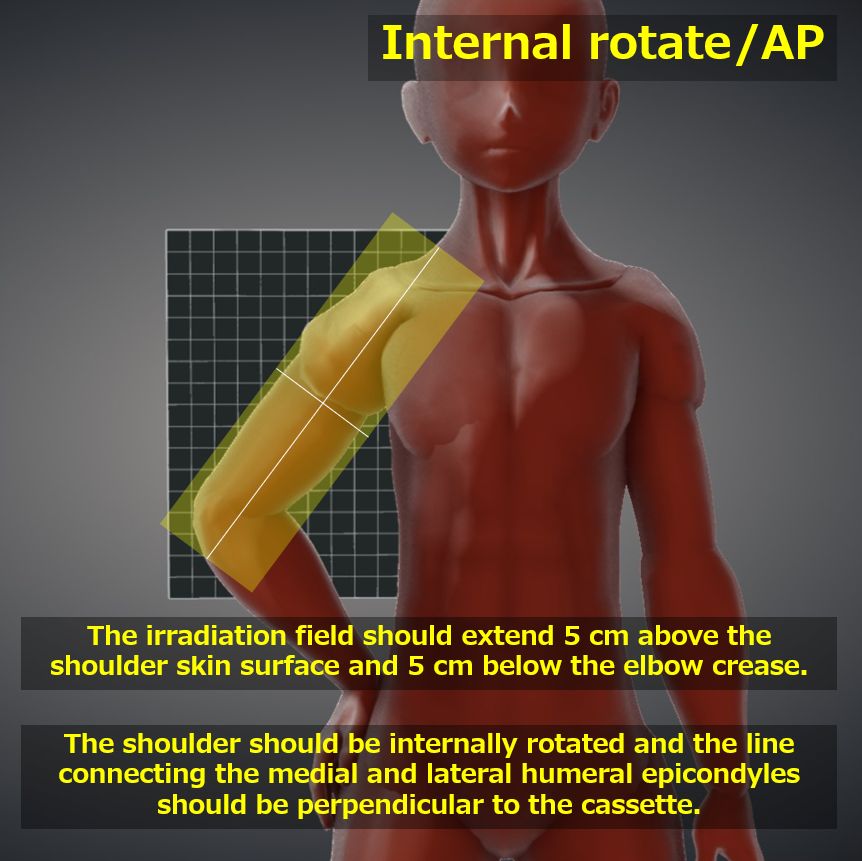
Both AP and PA directions can be used for imaging (see figure above).
The shoulder should be internally and externally rotated so that the elbow joint is lateral and the line connecting the medial and lateral humeral epicondyles is perpendicular to the cassette. Oblique position if necessary.
Select an appropriately sized cassette so that each shoulder joint to elbow joint is included with a margin of about 5 cm.
CR, distance, field size
CR : Vertical incidence at the left and right center of the upper arm at a height midway between the shoulder and elbow joints.
Distance : 100cm
Field size : The upper edge should be 5 cm above the shoulder skin surface, the lower edge should be 5 cm below the elbow crease, and the left and right sides should be limited to the skin surface.
Exposure condition
58kV / 10mAs
Grid ( – )
Suspend respiration.
Image, check-point
Normal (Radiopaedia)
Good contrast between the cortex and medulla of the humeral diaphysis should be observed.
Soft tissues should also be observable.
Shoulder to elbow joints should be included.
A lesser tubercle should be observed on the medial side of the humerus. The greater tubercle should overlap the humeral head.
The medial and lateral humeral epicondyles should overlap.
Videos
Related materials


