Purpose
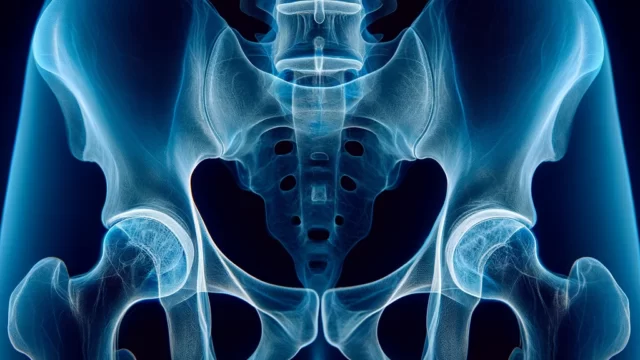
Observation of degeneration, fractures, and lesions of the acetabulum, femoral head and neck, greater trochanter, ilium, sciatic bone, and pubic bone.
Checking the status of metal in the body of patients who have undergone Total Hip Arthroplasty (THA).
Prior confirmation
Purpose of imaging, and whether or not THA surgery is indicated.
Presence or absence of suspected fracture. If a fracture is suspected, no internal rotation.
Remove obstacles.
Positioning
Lying on the back with no pelvic tilt (both anterior superior iliac spine equidistant from the bed).
Extend both lower limbs and rotate them inward (15-20°). Ensure that the patella is in a more internally rotated position than the middle-position.
With internal rotation, open the lower extremity so that the femoral axis is parallel to the sagittal plane.
If necessary, place weights on the ankle joint to fixate the posture.
Align the upper edge of the cassette with the upper edge of the iliac crest.
Align the center of the cassette with 2 transverse fingers head-side from the greater trochanter.
CR, distance, field size
CR : Intersection of the median sagittal plane and 4 cm (2 finger widths) cephalad from the greater trochanter.
Distance : 100cm
Field size : The area from the anterior superior iliac spine (and iliac crest if possible) to and including the proximal 1/3 of the femur. Left and right sides are restricted to the cutaneous surface. However, if the purpose is to check the status of metals in the body, all metals should be included.
Exposure condition
70kV / 20mAs
Grid ( + )
Suspend respiration during exposure.
Image, check-point
Patients with metals in the body (Radiopaedia)
The bilateral obturator foramen should be depicted symmetrically.
The THA and related instruments should be included.
Bony beams should be clearly visible.
Left and right markers should be included.
The femoral neck and greater trochanter should be extensively depicted.
The lesser trochanter overlaps the femoral stem.
The target area should have adequate contrast and tolerance.
Shenton lines should be observable.
Videos
Related materials
Garden classification ( Figure )
As one progresses from type I to IV, the likelihood of interrupted blood flow to the femoral head increases; in type III or IV, artificial head insertion is the treatment of choice.


