Purpose
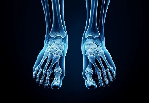
Observation of the longitudinal arch of the foot during weight-bearing in an upright position.
Diagnosis of flat feet.
Prior confirmation
Remove any obstacles.
Positioning
Place a positioning block of approximately 5cm on a stable, low platform and stand on it.
Position the cassette between both feet, touching the inside of the examined side foot.
Align the foot reference line (a line connecting the calcaneus and the second toe) of the examined side parallel to the cassette.
Apply natural (or maximal) weight-bearing on the examined side.
If there is a handrail for maintaining the posture, allow the patient to hold onto it.
Place the RL marker.
CR, distance, field size
CR : Horizontal incidence directed towards the intersection of a vertical line drawn down from the lateral malleolus and the sole of the foot.
Distance : 100cm
Field size : Narrowed down to include the range from the fingertips to the distal 1/3 of the lower leg bone.
Exposure condition
50kV / 5mAs
Grid ( – )
Image, check-point
Normal (Wikipedia)
Normal (musculoskeletalkey, Fig.3-9, Fig.3-33)
Ensure the measurability of the Calcaneal pitch and Tarso-first metatarsal angle.
Overlap between the inner and outer aspects of the talus.
Overlap of the proximal fibula with the distal tibia.
Observation of the calcaneus without overexposure.
Clear visibility of the cortex and trabeculae.
Presence of an RL marker.
No blurring due to movement.
Videos
Related materials


