Purpose
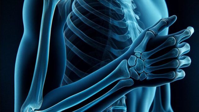
Observe the elbow joint from the front.
Fracture, dislocation, etc. (checklist)
Prior confirmation
Confirm the purpose of the radiography.
Confirm whether the purpose area is on the humeral or forearm side.
Positioning
Seat the patient in the chair.
Raise the upper arm, elbow joint, and wrist joint to shoulder height.
Extend the elbow joint. (Avoid hyperextension)
Slight external rotation so that the elbow joint is in frontage (the line connecting the medial and lateral epicondyles is parallel to the FPD).
If doing above position is difficult, tilt the patient’s entire body.
CR, distance, field size
CR : Distal by one finger lateral width at the center of the line connecting the medial and lateral epicondyles.
Distance : 100cm
Field size : Includes the half of humerus and forearm.
Exposure condition
50kV / 4mAs
Grid ( – )
Image, check-point
Normal (Radiopaedia)
Normal (Radiopaedia)
Abnormal (dislocation) (Radiopaedia)
Videos
Related materials


