Purpose
Diagnosis of femoroacetabular impingement (FAI). Measurement of the alpha angle.
Suitable for observing the femoral head and neck.
Excellent for observing the acetabulum, allowing confirmation of intra-articular foreign bodies.
Prior confirmation
Remove any obstacles.
Positioning
Supine position.
Flex the hip and knee joints of the side being examined to 90 degrees.
Abduct the lower limb of the side being examined by 30-45 degrees.
Use positioning blocks to prevent body movement.
Align the sagittal plane with the center axis of the cassette.
Align the X-ray center with the cassette center.
CR, distance, field size
CR : Perpendicular entry directed towards the center of the fold in the groin on the side being examined.
Distance : 100 cm.
Field size : Narrow down to approximately 15×15 cm, centered around the hip joint of the side being examined.
Exposure condition
75kV / 25mAs
Grid ( + )
Suspend respiration
Image, check-point
Normal
Fig.4 (B)
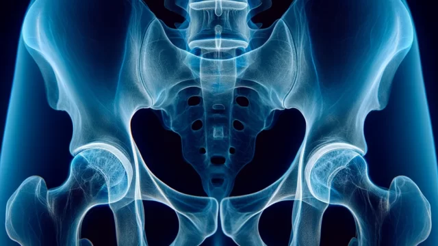
The femoral head, neck, greater trochanter, and acetabulum should be clearly visualized.
The alpha angle should be measurable. (Normal alpha angle is 50° or less)
There should be no motion blur due to movement.
Videos
Related materials


