Purpose
Project the relationship between the fetal head and maternal pelvis.
Measure the pelvic inlet transverse diameter and the interischial spine diameter.
Diagnosis of cephalopelvic disproportion (CPD) between the fetal head and maternal pelvis.
Although associated with less fetal exposure compared to the Multis technique, it is rarely performed in Japan. The original imaging involves bi-directional radiography.
Prior confirmation
Confirm patient’s understanding of radiation exposure.
Remove any obstacles.
Positioning
Supine position.
Slightly abduct and flex both knees.
Align the mid-sagittal plane with the center axis of the cassette.
Ensure no pelvic rotation; align both left and right anterior superior iliac spines equidistant from the cassette.
Place the measuring ruler parallel to the cassette, at the level of the greater trochanter.
Align the X-ray center with the cassette center.
CR, distance, field size
CR : Perpendicular incident directed towards a point 3 fingerwidths cranial from the greater trochanter on the mid-sagittal plane.
Distance : 100-130 cm.
Field size : Superior border includes iliac crests, inferior border includes ischial bones.
Exposure condition
100kV / 10mAs
Grid ( + )
Full expiration.
Image, check-point
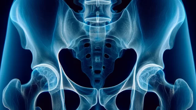
Ensure symmetrical projection of the obturator foramina on both sides.
Ensure symmetrical projection of the ischial spines on both sides.
Adequate contrast for measurement of inlet transverse diameter, midplane transverse diameter, and outlet transverse diameter.
Measuring ruler should be legible for readings of 10cm or more.
No motion blur due to movement.
Videos
Related materials
Literature opposing X-ray pelvimetry (1)
Literature opposing X-ray pelvimetry (2)


