Purpose
Observation of the middle talo-calcaneal joint, posterior talo-calcaneal joint, and tarsal sinus.
Prior confirmation
Remove any obstacles.
Positioning
Lateral decubitus position with the examined side down.
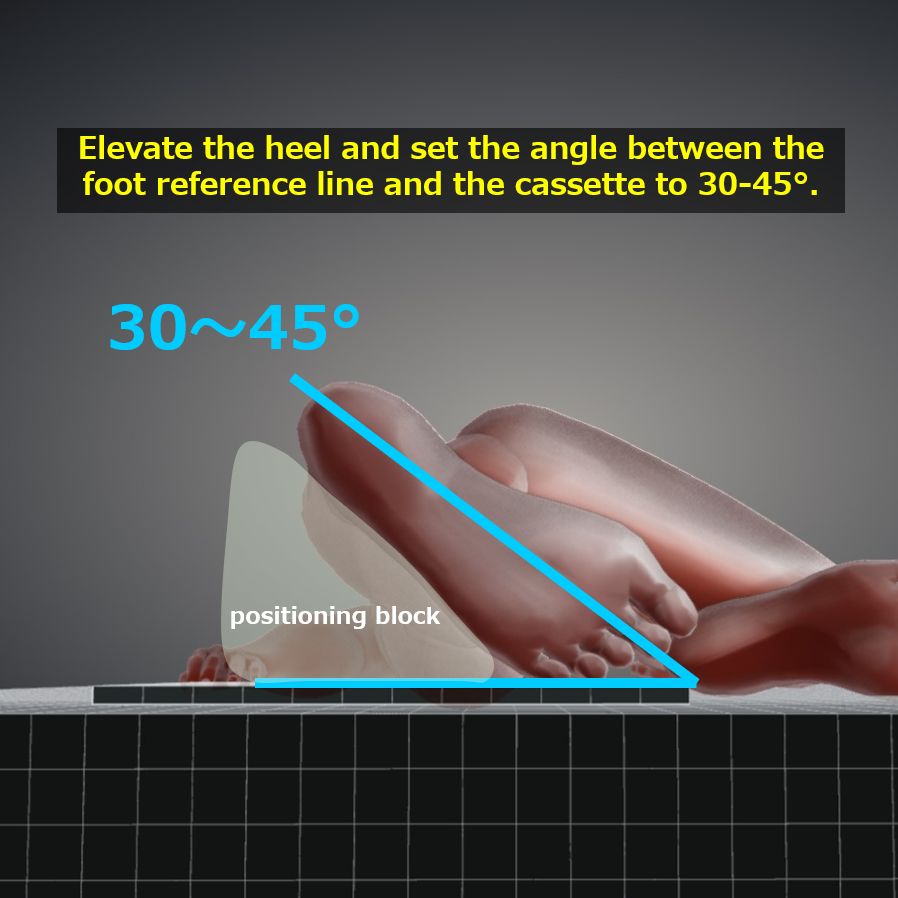
Position the non-examined lower limb slightly anterior to the examined side.
Align the sole of the foot parallel to the lower edge of the cassette.
Elevate the heel and set the angle between the foot reference line and the cassette to 30-45°.
Considering the oblique incidence at 20° in the head-to-foot direction, place the cassette on the foot side.
Place the RL marker.
CR, distance, field size
CR : Inferior from the medial malleolus by one fingerwidth, oblique incidence at 20° in the caudal direction.
Distance : 100cm
Field size : Narrowed down to include the area containing the calcaneus (approximately 15x15cm).
Exposure condition
55kV / 5mAs
Grid ( – )
Image, check-point
Normal (Fig.3)
Ensure that the posterior talo-calcaneal joint and the middle talo-calcaneal joint around the tarsal tunnel are consistently visible with uniform spacing.
The calcaneus is observable without overexposure.
Cortex and trabeculae are clearly visible.
There is sufficient tolerance to observe soft tissue.
Ensure the presence of an RL marker.
No blurring due to movement.
Videos
Related materials


