Purpose
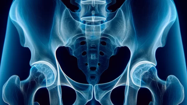
Supplementary AP or PA view of the abdomen is used to gain a three-dimensional understanding of the location and relationship of foreign bodies, tumors, calcifications, and abdominal aortic aneurysms.
Prior confirmation
Confirm the purpose of the imaging.
Adjust the field of radiation to 14×17 inches size.
Remove any obstacles (tubes, pads, buttons, etc.) that may interfere.
Positioning
Position the affected side in close contact with the cassette in an upright or lateral decubitus position (if unable to move, supine position with horizontal X-ray beam entry).
Align the center of the trunk with the central axis of the cassette.
Ensure the back is perpendicular to the cassette.
Elevate both arms to keep them out of the radiation field.
If in a lateral decubitus position, slightly bend the knees to stabilize the posture.
For upper abdominal imaging, align the upper edge of the radiation field with the lower edge of the scapula. For lower abdominal imaging, align the lower edge of the radiation field with the greater trochanter.
Place markers for erect / lateral decubitus position and RL orientation.
PositioninCR, distance, field size
CR : Perpendicular entry towards the center of the cassette.
Distance : 100-150 cm.
Field size : The irradiation fiels is 17 inches size in the vertical direction and narrowed to the body surface in the horizontal direction.
Exposure condition
80kV / 50mAs
Grid ( + )
Full expiration. (After complete exhalation, wait for 1 second before exposing the X-ray.)
Image, check-point
Normal (Radiopaedia)
When aiming for the upper abdomen, ensure that the left and right hemidiaphragms are projected.
When aiming for the lower abdomen, ensure that the pubic symphysis is projected.
The vertebral bodies near the center of the X-ray should be projected tangentially.
The edges of the gas pattern should be clear and there should be no blurring.
Videos
Related materials


