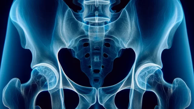
Sacrum Coccyx lateral view
Purpose
Observation of the sacrum and coccyx for the presence of fractures, dislocations, or signs of arthritis.
Prior confirmation
Remove any obstructing objects.
Positioning
Position the patient in the lateral decubitus position.
Align the longitudinal axis of the sacrum and coccyx with the central axis of the cassette.
Ensure that the mid-sagittal plane is parallel to the cassette. Use positioning blocks if necessary.
Keep the back perpendicular to the cassette to avoid any pelvic tilt.
Bend both knees to stabilize the posture.
CR, distance, field size
CR : Perform a perpendicular beam projection at a point 2.5 cm inferior to the anterior superior iliac spine and 3 cm from the posterior aspect.
Distance : 100cm
Field size : Narrow the field so that the upper border includes the iliac crests and the lower border reaches the level of the greater trochanters. The field should extend from the anterior edge of the L5 vertebral body to the posterior skin surface. Any areas protruding beyond the skin surface should be covered with a lead shield.
Exposure condition
80kV / 32mAs
Grid ( + )
Suspend respiration
Image, check-point
Fracture (Radiopaedia)
The pelvis and femoral heads should be clearly visible in accurate lateral view.
The entire sacrum and coccyx should be visible with sufficient contrast.
The lineae transversae of the sacrum should be identifiable.
L5 vertebra to the coccyx (3-5 segments) should be included.
The irradiation field should be minimized to the necessary extent.
Videos
Related materials


